Quantitative Muscle MRI Can Help Monitor Late-onset Pompe Disease, Study Shows


Magnetic resonance imaging (MRI) seems to be an effective and efficient tool to evaluate muscular status and monitor patients with late-onset Pompe disease (LOPD), according to researchers.
Their study, “Quantitative muscle MRI to follow up late onset Pompe patients: a prospective study,” was published in the journal Scientific Reports.
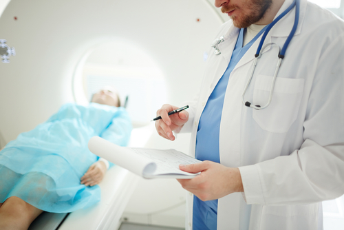
Several studies have demonstrated that quantitative muscle MRI is a valuable tool to monitor the progression of neuromuscular disorders. It a rapid, noninvasive diagnostic method that can assess the amount of fat, muscle tissue, and bone present in a specific spot. As a result, it can effectively determine the total muscle area and muscle tissue changes over time.
Results from a natural history trial (NCT01914536) conducted by a research team at Universitat Autònoma de Barcelona in Spain revealed that quantitative muscle MRI could help diagnose and assess muscle function in pre-symptomatic and symptomatic patients with Pompe disease.
To better understand the diagnostic and prognostic potential of muscle MRI the team followed 32 patients with LOPD for one year.
Twenty-two patients were symptomatic and treated with enzyme replacement therapies (ERTs). Ten patients were identified as nonsymptomatic as they did not have significant muscle weakness at clinical examination, but had elevated levels of creatine kinase (hyperCKemia) that suggested muscle damage.
The researchers found that at baseline (before the start of the study), symptomatic patients had, on average, about three times more fat in their thigh muscles than asymptomatic patients.
Also, those with higher amounts of thigh fat showed worse muscle function, as determined by the total Muscle Research Council (MRC) lower limb score, or results of the six-minute walk test (6MWT). No correlation was found between high fat fraction (FF) and respiratory parameters and patient-reported outcomes.
One year after the initial evaluation, a new assessment of muscle and respiratory function, as well as patient-reported outcomes, did not show significant changes in symptomatic patients. In contrast, MRI scans revealed a median increase of 1.7% in thigh muscle fat in this population.
No significant changes in thigh fat were reported in the asymptomatic LOPD patients group, or changes in muscle function after one year.
These findings demonstrated that quantitative MRI “was more sensitive to the changes over a period of one year than other more conventional muscle tests,” the researchers wrote.
They suggest that this evaluation method should be added “to the protocol currently used to follow up LOPD patients.” It could provide useful information to evaluate treatment response in symptomatic patients.
For asymptomatic patients, evaluation of fat infiltration detected by quantitative MRI “should be taken into account when considering ERT or a closer follow-up of progression,” they added.
Despite its diagnostic potential, the team highlights that use of quantitative MRI to effectively detect muscle degeneration is still debatable, and more studies are warranted.