Sugar marker found in urine could help track progression in LOPD: Study
Measuring Glc4 levels may help identify patients at higher risk of muscle decline
Written by |
Measuring levels of glucose tetrasaccharide (Glc4) in urine may offer a simple, noninvasive way to identify people with late-onset Pompe disease (LOPD) who are at higher risk of worsening muscle decline, even while receiving enzyme replacement therapy (ERT).
That’s according to a real-world analysis of 35 patients with LOPD who were followed up for four years.
Overall, these findings suggest Glc4 could serve as a “complementary biomarker for routine disease monitoring,” researchers wrote.
The study, “Urinary glucose tetrasaccharide tracks disease activity in late-onset Pompe disease,” was published in the journal Neuromuscular Disorders.

Monitoring disease progression a major challenge of managing LOPD
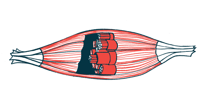
Pompe disease is caused by mutations in the GAA gene that lead to the absence or faulty production of acid alpha-glucosidase, an enzyme needed to break down a complex sugar molecule called glycogen. When this enzyme does not work properly, glycogen builds up inside cells — particularly muscle cells — interfering with normal muscle function.
In LOPD, symptoms develop after infancy and typically include worsening muscle weakness along with breathing difficulties.
Standard treatment for Pompe disease uses ERT, which provides patients with a lab-made version of the missing enzyme. Several ERTs are now available and can help improve or stabilize muscle strength, particularly if treatment is started early. However, treatment responses vary widely among patients.
One of the biggest challenges in managing LOPD is monitoring disease progression. Standard muscle strength and breathing tests may fail to detect subtle changes over short periods, making it difficult to assess how well treatment is working.
Researchers are therefore seeking reliable biomarkers — simple biological measures that more accurately reflect disease activity. One example is urinary Glc4, a glycogen breakdown product that increases when excess glycogen accumulates in the body. Glc4 is already used to monitor treatment response in infantile-onset Pompe disease, but its usefulness in LOPD is less well established.

Higher Glc4 levels tied to greater risk of worsening exercise capacity
In this study, a team led by Spanish researchers conducted a real-world analysis to evaluate whether urinary Glc4 levels could reliably track disease progression and treatment response in people with LOPD.
The analysis included patients with genetically confirmed LOPD who were enrolled in a four-year natural history study in Barcelona. Patients were grouped based on symptoms and treatment status, including symptomatic patients receiving ERT, symptomatic but untreated patients, and asymptomatic individuals. All treated patients received Myozyme (alglucosidase alfa, marketed in the U.S. as Lumizyme)
At the start of the study (baseline), urinary Glc4 levels varied widely among patients, with no significant differences observed between the groups. However, among symptomatic patients receiving ERT, Glc4 levels showed marked year-to-year fluctuations, with several patients experiencing changes greater than 10% relative to the previous year, “underscoring the need for longitudinal assessment rather than isolated measurements,” the team wrote.
While Glc4 levels did not correlate with muscle strength or MRI findings at individual time points, baseline Glc4 levels predicted long-term functional decline in symptomatic, ERT-treated participants. Higher Glc4 levels were associated with a greater risk of worsening exercise capacity over time, as assessed by the six-minute walk test.
[These findings suggest that] sustained high Glc4 may be associated with greater risk of progression, and therefore Glc4 could serve as a complementary biomarker to support clinical decision-making regarding initiation or modification of ERT.
Further analysis identified a baseline Glc4 cutoff of about 13 mmol/mol creatinine that helped distinguish patients at higher risk of motor deterioration. Symptomatic patients on ERT whose Glc4 levels were higher than this threshold experienced, on average, a substantial decline in walking distance, reduced motor function, and greater fat replacement in thigh muscles over three years.
Importantly, higher baseline Glc4 levels remained a strong predictor of functional decline, even after accounting for age, baseline walking ability, and time on treatment.
Among asymptomatic patients, Glc4 levels followed variable patterns over time. Only one individual, who showed a progressive increase in Glc4 levels, developed mild muscle symptoms during follow-up. In several patients who began ERT during the study, Glc4 levels initially decreased after the start of treatment, although long-term trends varied.
“All asymptomatic patients had Glc4 levels above normal, indicating that glycogen accumulation occurs even before overt symptoms,” the investigators wrote. “This observation is consistent with data from newborn screening programs, where infants identified with LOPD frequently show persistently elevated Glc4 levels despite remaining clinically stable for years.”
Together, these findings suggest that “sustained high Glc4 may be associated with greater risk of progression, and therefore Glc4 could serve as a complementary biomarker to support clinical decision-making regarding initiation or modification of ERT,” the scientists concluded.
Study limitations noted by the researchers include its relatively small number of participants and that Glc4 levels were measured “without dietary, activity, or timing standardization, introducing potential variability unrelated to disease activity.”
“These findings derive from a single-center cohort and require validation in larger, independent populations,” they noted.