Muscle MRI scans show promise for monitoring LOPD: Study
Pattern changes correspond to strength, mobility measures
Written by |

Quantitative MRI (qMRI) imaging of the muscles is a useful technique for identifying signs of compromised muscle health in people with neuromuscular conditions like Pompe disease, according to a small study.
The study identified specific patterns of qMRI changes in the thigh muscles of people with late-onset Pompe disease (LOPD) that correlated with clinical measures of muscle strength and mobility. Patterns observed in LOPD were distinct from other neuromuscular conditions.
“Potential future uses of qMRI may encompass disease monitoring, prognosis assessment, participation in clinical trials, and selection of treatment modalities,” the researchers wrote. The study, “Evaluation of Neuromuscular Diseases and Complaints by Quantitative Muscle MRI,” was published in the Journal of Clinical Medicine.
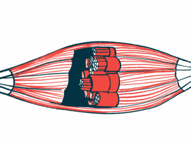
Neuromuscular disease (NMD) encompasses a group of hundreds of conditions wherein muscle weakness arises due to problems with the nerves that control muscles, the muscles themselves, or the sites where nerves and muscles communicate, called the neuromuscular junction.
Among them is Pompe disease, in which a large sugar molecule abnormally accumulates inside cells because the enzyme that normally breaks it down is lacking. Muscles are particularly susceptible to this accumulation, leading to symptoms of progressive muscle weakness.

Direct comparison difficult
qMRI is a way of evaluating and monitoring muscle health across different types of NMD. It can capture muscle wasting, fat infiltration into the muscles, structural changes, and inflammation, among other aspects of muscle health. It has the ability to see subtle signs of disease progression that may not yet be clinically apparent.
However, it is not always easy to directly compare qMRI findings from different studies and across different conditions because there aren’t any standardized protocols for the analyses.
In their study, the researchers used a standard qMRI protocol to look at aspects of muscle health in 10 people with LOPD as well as individuals with other forms of neuromuscular involvement and healthy people, who served as a control group.
Other conditions included limb-girdle muscular dystrophy (LGMD) and inclusion body myositis (IBM) — both rare NMDs — and people with neuromuscular complaints after a COVID-19 infection.
Results showed that the proportion of fat in the muscles was significantly higher in people with LOPD relative to controls, which was similarly observed in LGMD and IBM, but not in post-COVID individuals.
The infiltration of fat into muscle tissue is reflective of lower muscle quality, and is commonly observed in neuromuscular diseases, including LOPD.
In LOPD patients, the hamstring muscles at the back of the thigh were largely affected, with others in the legs relatively spared. And a higher fat fraction in the thigh muscles correlated with worse performance on a clinician-rated measure of muscle strength and lower mobility in LOPD patients.
“The ability to detect subtle changes in muscle fat content using qMRI holds significant promise for early diagnosis and precise monitoring of disease progression and treatment efficacy,” the researchers wrote.
Still, once fat replacement is observed, it is not reversible, according to the authors. Other qMRI metrics might be able to capture earlier changes in muscle health before that process sets in.
One such metric is called water T2 relaxation time, an MRI measure related to water content in the tissue. It has been proposed as a biomarker for muscle diseases, since it increases with things like swelling, inflammation, and muscle damage.
Indeed, both LOPD and IBM patients exhibited an elevated water T2 relaxation time compared with controls. While the measure could offer a biomarker of disease activity in these conditions, the researchers noted that it is not specific to any one disease process.
Other measures evaluated in the muscle MRI scans included diffusion metrics, which broadly look at how water molecules move in muscle tissue. This can tell researchers something about structural changes in the muscles.
All of the NMDs, as well as post-COVID patients, showed a significantly elevated diffusion metric called fractional anisotropy. In LOPD patients, diffusion metrics in the thigh correlated with clinical measures of muscle strength.
When the findings are taken together, “qMRI emerges as a helpful marker in the assessment of NMD,” the researchers wrote.
Still, “standardised and simplified imaging protocols and data analysis are needed to ensure the broad deployment of qMRI techniques and comparability between different centres,” they wrote.