
Chest MRI May Capture Respiratory Muscle Weakness at Early Stages
Written by |
A chest MRI can be used to detect early signs of respiratory muscle weakness in people with Pompe disease, even when lung function is still within a normal range, a study reported.
According to its investigators, the findings highlight the importance of these imaging tests to rapidly identify patients in need of treatments that might prevent irreversible damage to their respiratory muscles.
The study, “Chest MRI to diagnose early diaphragmatic weakness in Pompe disease,” was published in the Orphanet Journal of Rare Diseases.
Breathing difficulties caused by respiratory muscle weakness are commonly observed in patients with diseases that compromise muscle health, as is the case of Pompe.
In these people, particularly patients at more advanced disease stages, weakness of the diaphragm — the large dome-shaped muscle that allows the chest to move with each breath — is the main cause of respiratory dysfunction.
Although certain measures of lung function that are captured with spirometry usually reflect changes in the diaphragm, mild impairments may not be picked up by these tests.
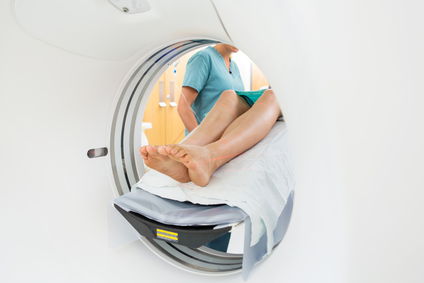
Investigators in the Netherlands explored the usefulness of chest MRI scans at detecting early signs of diaphragm weakness in people with Pompe.
A total of 35 patients, ages 15–70, who visited the Center for Lysosomal and Metabolic Diseases at the Erasmus MC University Medical Center between 2016 and 2018, were included in their study. Patients were matched by sex and age with 18 healthy individuals, who served as controls.
Spirometer-controlled MRI scans were given to all study participants, and used to assess the motion and shape of the diaphragm while participants breathed in and out.
Conventional spirometry tests were also used to measure forced vital capacity (FVC), which assesses lung health by measuring the amount of air a person is able to exhale after a deep breath. FVC was measured while participants were in upright seated and supine positions before their MRI scans.
Thirteen Pompe patients had normal spirometry results (FVC values of 80% or greater while in a supine position), while the other 22 had abnormal results, meaning lower supine FVC values.
A comparison of these patient groups found those with normal FVC values were younger (mean age of 31 vs. 45), had a shorter disease duration, and fewer of them had been given enzyme replacement therapy — 46 vs. 82%. As expected, FVC values were higher in the control group.
Analysis of diaphragm motion captured on the MRI scans showed a lesser degree of muscle up and down movement with each breath in patients with abnormally low FVC values, compared with patients with normal FVC values and with healthy controls.
No differences were evident among the three groups when regarding the diaphragm’s ability to move back and forth with each breath.
But the diaphragm of patients took on a more marked curve shape during inhalations, relative to what was seen in controls — with a significantly more accentuated curvature evident in patients with abnormal spirometry results.
“We found that motion of the diaphragm is decreased in patients with Pompe disease, and that the curvature of the diaphragm in these patients is increased during inspiration. Interestingly, these MRI signs were observed even in Pompe patients whose spirometry results were still within the normal range,” the researchers wrote.
“We hypothesize that the aberrant shape of the diaphragm during inspiration is caused by diaphragmatic weakness,” they added.
They concluded that “MRI is a sensitive tool for detecting early stages of diaphragmatic weakness in patients with Pompe disease,” including those with normal spirometry measures. Use of this tool “might help to select patients for early intervention to prevent possible irreversible damage to the diaphragm,” the researchers noted.


Leave a comment
Fill in the required fields to post. Your email address will not be published.