Muscle MRI Reveals Deterioration in Late-onset Pompe Patients, Study Says
Written by |

People with late-onset Pompe disease (LOPD) show significant increases in the amount of muscle replaced by fat while on treatment, which is linked to reduced muscle strength and motor function, according to a new study.
These findings support the use of muscle imaging to assess disease progression in clinical trials, the researchers said.
The study, “Follow‐up of late‐onset Pompe disease patients with muscle magnetic resonance imaging reveals increase in fat replacement in skeletal muscles.” was published in the Journal of Cachexia, Sarcopenia and Muscle.
In many cases, the first symptom of LOPD is muscle weakness, especially in the torso and legs. As in other diseases involving muscle deterioration, fat replaces muscle tissue in people with Pompe disease.
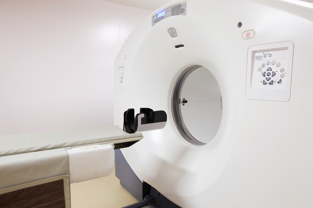
MRI is seen as a valuable tool for both diagnosis and follow-up of patients with muscle disorders. A specific imaging technique known as the Dixon MRI sequence can measure the amount of fat present in skeletal muscle.
When applied in two large studies with LOPD patients, Dixon MRI was able to identify and measure an increase in fat replacement over one year.
However, these studies did not assess the change in fat replacement over time or identify which demographic or clinical features may influence this progression.
To address this knowledge gap, researchers at Universitat Autònoma de Barcelona, in Spain, used Dixon MRI to observe the changes in fat replacement in skeletal muscles of LOPD patients over four years.
The open-label, Sanofi Genzyme-funded study (NCT01914536) involved 36 people with LOPD (average age 43.9). Patients were evaluated during four annual visits. At each visit, tests for muscle and lung function (using spirometry) were performed, along with the quantitative Dixon MRI of muscles and quality of life assessments.
At the beginning of the study, 23 participants were being treated over approximately four years with Sanofi Genzyme’s Myozyme (alglucosidase alfa, also sold as Lumizyme in the U.S.), a type of enzyme replacement therapy.
Of the 13 remaining patients, nine remained pre-symptomatic throughout the study period, and four started treatment with Myozyme during follow-up due to the onset of muscle weakness. As such, they were excluded from the statistical analyses.
Results showed that patients experiencing symptoms had an increased fat fraction (5.7%) in the thigh by the end of follow-up. This corresponded to an increase of 1.9% per year. Despite significant variability from one patient to another, almost all muscles analyzed showed this increase in fat fraction.
A smaller, yet significant, increase (2.6%, 0.8% per year) was found in thigh muscles of pre‐symptomatic patients.
However, in some pre‐symptomatic patients, the research team found higher fat fractions in a hip muscle (adductor major) and in muscles of the back (paraspinal).
The team found that older age was associated with higher fat fraction in these muscles in pre-symptomatic patients. Also in these participants, those older than 25 showed increasing fat fraction in the thigh over follow-up. No such change was seen in younger patients.
Then, the investigators found that increased fat fraction in the thigh was associated with lower muscle strength, most notably the strength to extend the knee. This was measured by hand‐held myometry and the Medical Research Council (MRC) scale.
Significant correlations also were identified between higher thigh fat fraction and worse performance in motor tests — such as the time to walk 10 meters (33 feet) and the six minute walk test.
Early onset of symptoms, longer treatment periods, and both muscle function and fat fraction at baseline were all significantly linked to a greater increase in fraction at follow-up.
Finally, to identify which specific muscles to measure to follow fat replacement over time, the patients were divided into four groups depending on their mean thigh fat fraction at baseline.
The first group included nine pre‐symptomatic patients, who already had high values of fat fraction in paraspinal muscles at baseline. The second group of 11 symptomatic patients showed highest fat fraction in both paraspinal and adductor major.
In turn, the third group of six patients and the fourth group of 11 participants, all symptomatic, had large fat replacement in several muscles. A thigh muscle known as the vastus lateralis was the best candidate to monitor fat replacement in these patients.
“Our study identifies that skeletal muscle fat fraction continues to increase in patients with LOPD despite the treatment with enzymatic replacement therapy,” the researchers wrote.
“Hereby, we show that fat fraction along with muscle function tests can be considered a good outcome measures for clinical trials in LOPD patients,” they added.


