
New Ultrasound Parameter May Help Assess Muscle Changes
Researchers examine echo heterogeneity index in patients' ultrasounds
Written by |

The level of variability seen during an ultrasound — a parameter called echo heterogeneity index — may help to quantify the level of skeletal muscle involvement in Pompe disease patients, a new study suggests.
A lower EH index in the leg muscles was indicative of worse motor function, as shown by impaired walking and stair climbing.
The study, “Sonographic Evaluations of the Skeletal Muscles in Patients with Pompe Disease,” was published in the European Journal of Paediatric Neurology.
Pompe disease is caused by mutations in the GAA gene, which provides instructions for making an enzyme called acid alpha-glucosidase (GAA). This enzyme is responsible for the breakdown of glycogen — a sugar molecule that serves as energy storage.

Despite the availability of enzyme replacement therapy (ERT), the mainstay and only therapeutic approach approved for Pompe, most patients still experience muscle weakness, especially in muscle fibers that already were severely damaged or accumulated substantial glycogen before starting therapy.
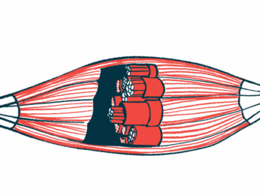
Ultrasounds — a noninvasive imaging test — are used to assess muscle involvement in Pompe disease patients before the onset of muscle weakness signs. The Heckmatt scale is a four-point tool that uses ultrasound to evaluate muscle damage. Specifically, it assesses muscle echogenicity, the ability to reflect or transmit ultrasound waves, as a measure of the amount of fat and scarring. Increasing scores indicate greater fat infiltration and scarring.
However, among other limitations, the Heckmatt scale grading is subjective. As such, more objective and quantitative ultrasound-measuring tools to monitor muscles changes and motor function in Pompe disease are still lacking.
The EH index
In this study, researchers in Taiwan tested two ultrasound parameters: shear modulus and the echo heterogeneity (EH) index. The shear modulus is used to measure tissue stiffness, while the EH index is a new indicator of muscle changes. Typically, a healthy muscle has a more heterogenous echogenic appearance (lower Heckmatt score) and a high EH index, but once a muscle becomes damaged and scarred (higher Heckmatt score), the EH index decreases.
“We aimed to find out a ultrasonographic parameter that can quantify the muscle involvement and correlate with the motor functions in Pompe disease,” the researchers wrote.
To test the two parameters, the researchers performed ultrasound imaging in four leg muscles (rectus femoris, biceps femoris, tibialis anterior, and medial gastrocnemius), and two arm muscles — biceps brachii and triceps brachii. Ultrasounds were performed in the non-dominant limbs, because dominant limbs are more likely to present overuse injuries.
Following the ultrasounds, three functional tests were performed. These included muscle power, the six-minute walk test — the maximum distance an individual is able to walk over six minutes on a hard, flat surface — and the four-step stair climb. All tests were perfomred by a physical therapist, who was blinded (unaware) of the ultrasounds’ results.
If the patient was tired after imaging, functional tests were completed on another day within one month.
Patient profiles
In total, 18 Pompe disease patients (six females and 12 males, median age 13.1 years) participated in the study. Ten patients had infantile-onset Pompe disease (IOPD), and seven had late-onset Pompe disease (LOPD). One had an unknown type, which was diagnosed during newborn screening (no symptoms were visible at the time of the study).
Among the 18 patients, 16 received ERT. Those with IOPD were treated for a median of 9.9 years and LOPD patients for 10.4 years. Five participants (three males and two females) didn’t perform the functional tests.
Results showed that the EH index was inversely proportional to the Heckmatt scale grade, meaning that when the EH index increased, the Heckmatt score decreased.
A moderate negative correlation — meaning the greater one, the lower the other — was seen between the EH index and Heckmatt score in the biceps femoris muscle. A strong negative correlation was seen in the five other muscles.
The shear modulus showed a significant positive correlation — meaning the greater one, the greater the other — with the Heckmatt score for the biceps femoris muscle, while a negative correlation was found for the triceps branchii. No other significant correlations were observed in other muscles.
The EH index of the six evaluated muscles were lower in participants with classical IOPD compared to LOPD, in particular for the tibialis anterior, medial gastrocnemius, and the biceps brachii.
In the LOPD group, the rectus femoris was the most affected muscle, while the triceps brachia was the least and last muscle to show changes.
Although muscle strength was not correlated with EH index values, the walking distance in the six-minute walk test showed a moderate positive association with the EH index of the tibialis anterior and medial gastrocnemius muscles.
The duration of the four-step stair climbing was negatively correlated with the EH indiex of the leg muscles, meaning that a patient taking longer to climb four stairs showed a lower EH index.
Quantifying muscle involvement
Overall, these findings suggest that the EH index may be used to quantify muscle involvement in Pompe disease patients, whereby lower indices in the muscle legs reflect worse motor function.
Also, “the EH index can be adopted as a US [ultrasound] evaluation approach to monitor disease severity and assess ERT response,” the researchers wrote.
“Further studies with larger samples … will be warranted in patients with Pompe disease,” they concluded.

Leave a comment
Fill in the required fields to post. Your email address will not be published.