The role of muscle MRI in tracking Pompe disease
 Fact-checked by
 Pompe disease is a genetic condition that causes progressive muscle weakness, along with other symptoms. Muscle MRI scans are a noninvasive imaging method that creates detailed images of muscles and helps monitor disease progression.
Beyond diagnosis, proactive Pompe disease MRI monitoring helps doctors and patients better understand how the disease is evolving and supports informed treatment decisions.
Pompe disease muscle MRI can sometimes detect changes in muscle structure before symptoms appear, especially in people with late-onset Pompe disease (LOPD). Muscle imaging in Pompe disease can also help doctors track responses to treatment.
Why monitoring muscle health matters
Pompe disease is caused by mutations in the GAA gene, which provides instructions for making the acid alpha-glucosidase (GAA) enzyme. Without enough functional GAA, glycogen — a complex sugar — builds up inside cells and causes damage.
Pompe disease affects muscles in particular because muscle cells normally rely on glycogen for energy. When glycogen accumulates, muscle cells are damaged and deprived of their main energy source, leading to progressive weakness and muscle wasting. Over time, healthy muscle tissue is replaced by fat.
Muscles are resilient and can continue to function even when some fibers are damaged. As a result, muscle damage in Pompe disease may progress silently for a period of time without obvious symptoms, particularly in LOPD.
For this reason, monitoring muscle damage in Pompe disease using noninvasive imaging, such as muscle MRI, is important for tracking disease progression. MRI findings may also help predict complications, including breathing problems related to respiratory muscle weakness.
What muscle MRIs show
Muscle MRI may show characteristic patterns of muscle damage in people with Pompe disease, including:
- muscle wasting, or atrophy
- fat infiltration
- increased water content, reflecting areas of active damage
These MRI findings in Pompe disease are most pronounced in people with late-onset disease. In infantile-onset Pompe disease, muscle weakness and glycogen buildup occur more rapidly, and fat infiltration is less prominent.
Unlike some other neuromuscular conditions, muscle inflammation is not typically a major feature seen on MRI in Pompe disease.
When monitoring Pompe disease with MRI, doctors often see increasing muscle damage and fat accumulation over time, which tend to correlate with declining muscle function. Additional muscle groups may become affected as the disease progresses.
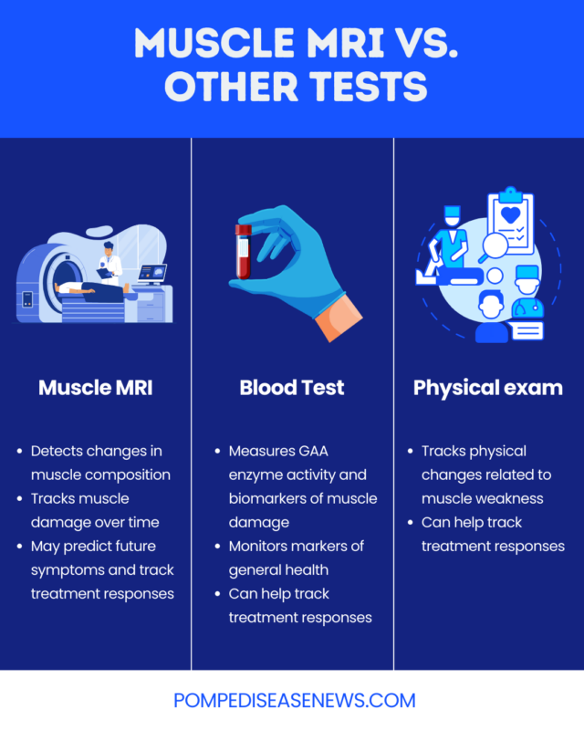
Muscle MRI is useful for showing structural changes, but it cannot measure how well a muscle is functioning, and standard scans typically do not show the amount of glycogen that has accumulated. Using other tests, such as blood tests and physical examinations, alongside muscle MRI for Pompe disease progression monitoring can provide a clearer overall picture of disease status.
How muscle MRI helps track disease progression
Using muscle MRI for Pompe disease progression tracking offers several potential advantages, including:
- helping identify subtle changes before symptoms emerge or worsen
- enabling planning of treatment and physical therapy efforts
- monitoring stability or progression over time
- objectively measuring disease activity
Alongside functional assessments of breathing abilities, muscle MRI may also play a role in monitoring respiratory problems related to muscle weakness in Pompe disease.
Muscle MRI and treatment monitoring
Muscle MRIs can also help with Pompe disease treatment monitoring.
Enzyme replacement therapy (ERT), which provides the body with a working version of GAA to support glycogen breakdown, is the primary Pompe disease treatment approach. Although ERT does not usually stop fat from infiltrating muscles, it may slow these changes and help preserve muscle function, effects that can be seen on muscle MRI.
Doctors also typically track responses to ERT in other ways, including with physical exams and blood tests, as part of Pompe disease follow-up care.
What to expect during a muscle MRI scan
Before an MRI, patients will be asked to remove any metal items, such as jewelry and watches.
During the appointment, an MRI technologist will position the patient on an exam table that slides into a tunnel-shaped scanner. The scanner makes loud noises while images are taken, and the person must remain very still. A muscle MRI typically lasts about 30 to 45 minutes.
Some MRI scans use contrast agents to help certain structures appear more clearly. For these scans, a technologist injects the contrast agent into a vein before imaging begins.
Some people may feel uncomfortable or anxious inside the enclosed MRI scanner. To help with comfort during the scan, people may use relaxation techniques and other strategies, such as:
- wearing an eye mask or blanket
- listening to music through provided headphones or speakers
- practicing deep breathing or meditation
For people with significant anxiety, a doctor may prescribe a mild sedative before the exam. Those with Pompe-related muscle weakness or breathing difficulties should discuss the safest comfort options with their healthcare provider.
How often are muscle MRIs used in Pompe disease?
There is no consistent clinical consensus on how often muscle MRI and other imaging tests should be performed for people living with Pompe disease. Frequency may depend on:
- disease type
- treatment regimens
- symptoms and their progression
The benefits of regular MRI monitoring are best established for people with late-onset Pompe disease, who tend to show clearer muscle changes. For these individuals, muscle MRIs may be recommended every one to two years, though recommendations may change over time.
Pompe Disease News is strictly a news and information website about the disease. It does not provide medical advice, diagnosis, or treatment. This content is not intended to be a substitute for professional medical advice, diagnosis, or treatment. Always seek the advice of your physician or other qualified health provider with any questions you may have regarding a medical condition. Never disregard professional medical advice or delay in seeking it because of something you have read on this website.