
Study maps molecular changes in Pompe muscle cells
Muscle cells show dysregulation across range of biological processes
Written by |
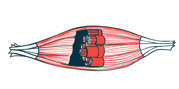
Muscle cells in people with Pompe disease show dysregulation affecting a range of different biological processes, including molecular recycling systems, muscle fiber structure, immune activity and metabolism, according to a study.
Specific patterns of molecular dysregulation were generally more pronounced in muscle from people with infantile-onset Pompe disease (IOPD) than late-onset Pompe disease (LOPD), aligning with differences in clinical severity. Results also indicated that treatment with enzyme replacement therapy (ERT) can help to normalize the molecular activity within muscles in IOPD, similar to what has been previously reported in LOPD.
The study, “Identifying regulatory muscle processes in infantile and adult Pompe patients using a multi-omic and morphological approach,” was published in Brain Pathology.

Muscle biology
Pompe disease is caused by mutations in the gene that provides instructions to make the acid alpha-glucosidase enzyme (GAA). This enzyme is normally needed to break down glycogen, a complex sugar; in Pompe, the enzyme is absent or dysfunctional, so glycogen cannot be broken down, and it builds to toxic levels in cells. Muscle cells, which normally use glycogen to store sugar for fuel, are especially vulnerable to damage in Pompe.
The two main types of Pompe are IOPD, in which symptoms appear during infancy, and LOPD, in which symptoms appear later in childhood or in adulthood. There can be considerable variability in how Pompe manifests from person to person, but in general, IOPD is more severe than LOPD.
Although it’s broadly understood that muscle damage in Pompe is driven by toxic glycogen buildup due to GAA deficiency, the details of how muscle biology is altered in Pompe remain incompletely understood. It’s also not clear to what extent muscle biology differs in IOPD and LOPD.
To gain more insight, a team of scientists in Germany conducted a series of analyses using muscle biopsies from 21 people with LOPD and 11 people with IOPD, none of whom had received treatment. The analyses included morphological assessments — that is, analyses of the physical structure of muscle cells — as well as proteomics and transcriptomics, which assess total protein levels and gene activity in the cell, respectively.
“Integrating proteomics with transcriptomics provided a multi systems-level view of [gene activity] and protein networks simultaneously, offering a more comprehensive overview in Pompe disease than examining either layer individually,” the researchers wrote.
Data from the Pompe samples were compared against muscle biopsies from similarly aged individuals without muscle disease. Results showed that, in Pompe disease, muscle cells show dysregulation across a range of biological processes.
For example, the researchers found that in both types of Pompe, muscle cells show abnormalities in autophagy, a form of molecular recycling that cells use to break down unneeded molecules into simple components that can be repurposed. Both forms of Pompe disease were also marked by increased activity of genes related to immune system activation and abnormalities related to the sarcomere, a specialized molecular structure that muscle cells use to help power muscle contractions.
The researchers noted that these biological abnormalities were generally more pronounced in samples from people with IOPD than in LOPD, which was expected given that IOPD is generally more severe clinically.
“Consistent with the … clinical presentation, these data indicated stronger differences using transcriptomic and proteomic analysis in IOPD compared to controls than in LOPD,” they wrote. “Additionally, LOPD samples showed some overlap with control samples, which is consistent with the finding that some muscle biopsies present with [less damage in LOPD].”
ERT is an approved Pompe treatment designed to deliver a working form of the GAA enzyme. Previous studies have suggested that ERT can help normalize molecular activity in muscle cells from LOPD patients, but it hasn’t been clear if similar effects are also seen in IOPD. In an additional analysis, the researchers looked at muscle biopsies from five IOPD patients who’d been on ERT, and results showed changes similar to what’s been previously reported in LOPD.
“Our data show that ERT has a positive effect on the function of skeletal muscles in IOPD, including the upregulation of developmental, sarcomeric, immune system and metabolic processes,” the scientists concluded.



Leave a comment
Fill in the required fields to post. Your email address will not be published.