
Severe Impairments Seen in Diaphragm Motion, Study Shows
Written by |

Pompe disease patients show severe impairments in diaphragm motion, the major muscle of respiration, when compared with those who have other muscle disorders or motor neuron diseases, a small study reports.
The study, “Diaphragmatic dysfunction in neuromuscular disease, an MRI study,” was published in the journal Neuromuscular Disorders.
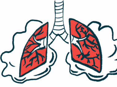
Respiratory insufficiency is a common problem among people with Pompe disease or other neuromuscular diseases. It is often caused by weakness of the diaphragm, the major muscle of respiration that sits below the lungs and separates the chest from the abdomen. Weakness in additional respiratory muscles also plays a role.

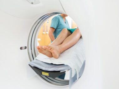
Evaluation of respiratory muscles could be informative about disease course and how patients may respond to therapy. While spirometry is the most common test for evaluating overall pulmonary function, MRI scans can assess the function of specific respiratory muscles, like the diaphragm.
A recent study has shown that MRI can also be used to investigate early diaphragm impairment in Pompe disease patients. However, whether these deficits are also present in other neuromuscular diseases or are specific to Pompe patients is unclear.
In this exploratory study, a team led by researchers in Rotterdam, Netherlands, used MRI and spirometry tests to evaluate diaphragm function relative to other muscles involved in breathing — called intercostal muscles, located between the ribs — across people with different neuromuscular diseases and with respiratory muscle weakness.
Their analysis included 35 adult Pompe patients (mean age 40) and 12 patients with a different neuromuscular disease. Seven of those had a clinical disorder of the skeletal muscles (a group of diseases called myopathies, mean age 51). Five had some form of motor neuron disease (mean age 67), a group of progressive neurological disorders that destroy nerve cells that control muscle control (three had progressive spinal muscular atrophy and two amyotrophic lateral sclerosis). Eighteen healthy participants were included to serve as controls.
Pulmonary function assessments included forced vital capacity (FVC) — it measures the amount of air that can be forcibly exhaled from the lungs after taking a deep breath — conducted while in an upright seated or supine position (lying horizontally with the face and torso facing up). Measures of respiratory muscle strength included maximum static inspiratory pressure (MIP) and expiratory pressure (MEP) in the upright position.
FVC of Pompe patients showed no significant differences to participants with myopathies or motor neuron diseases. These two groups — whose participants were older and more often male and active smokers — performed worse than healthy controls.
Diaphragm weakness, however, was significantly higher among Pompe patients when compared with people who have myopathies, 15% vs. 3%, according to the postural drop in FVC when moving from an upright position to a supine position.
MEP was significantly lower in patients with myopathies than in those with Pompe (mean 54% vs. 80%). No differences were seen regarding participants with motor neuron diseases.
In MRI scans conducted when patients breathe in and the diaphragm tightens, moving down toward the abdomen, results showed that this contraction was smaller in patients with myopathies than in healthy controls, but not different than in Pompe patients. No differences were seen across any of the groups regarding the motion of the overall thoracic muscles.
Compared with healthy controls, patients with myopathies or Pompe disease showed signs of insufficient diaphragmatic contraction.
However, when analyzing the diaphragm motion relative to the thoracic muscles, the researchers found that diaphragm motion was particularly impaired in Pompe patients.
A statistical analysis then revealed a correlation between pulmonary function and MRI outcomes.
“We conclude that spirometry-controlled MRI enables us to investigate respiratory dysfunction across neuromuscular diseases, suggesting that the diaphragm is affected in a different way in myopathies and motor neuron diseases,” the scientists wrote.
“Whether MRI can also be used to evaluate progression of diaphragmatic dysfunction requires additional studies,” they added.

Leave a comment
Fill in the required fields to post. Your email address will not be published.